ShareThis

Wednesday, January 11, 2023

Honey Bee vaccination: good, ugly or bad?


The less than saintly honey bee (copyright 2023 Emerald Biology)
 

For years I've been a distant proponent of the honey bee, thinking it a much beloved native species that was essential to plants and putting the natural healthy sweetner on my table. The delightful glow of going to the farmers market and picking up local honey.

A few months ago, I read that actually honey bees are not native to the United States but are in fact a European import (1). Not only that but they compete with native species for food, spread disease, and because beehives are moved around, the honey bees get an advantage over native species. This article in the Scientific American (2) explains the complexity of the situation admirably.

I'm all for the elimination of the use of pesticides and antibiotics; a reduction in the addition of chemicals to the environment can only help all species but the introduction of a vaccine (3) to deal with foulbrood in honey bees seems to be a step that helps commercial interests much more than keeping the native ecosystem in balance. I feel righteous indignation in my pure minded eco warrior stance. BUT I also read that honey bees are essential for agriculture to achieve the level of production need to feed the population at reasonable cost. I've already seen my grocery bill increase. Can I afford my righteous indignation? What would be the consequences of not using the vaccine and looking at ways to promote native pollinators? Can we do without honey?

It turns out that there are 4000 species of bees (4) and there are plants they prefer. npr (5) suggests a number of ways to support native species. Can agriculture do the same and still be commercially viable?


Tuesday, May 10, 2022

Lower and faster

Great news about a enzymes that degrade plastic (of a certain type) faster at lower temperatures: see it here

Sunday, April 7, 2019

Fuels for Biofuels part 6: the cellulosome

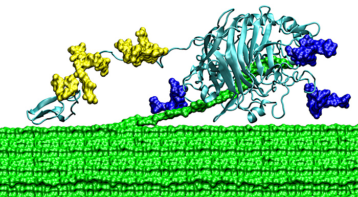
In the last post (yes, its been a while!) I talked about free cellulases. The problem with free cellulases is recovery and that you need several different types of cellulases to work together to accomplish the breakdown of cellulose. What if you could have a scaffold that did that for you? Bacteria and fungi have done just that and its called the cellulosome! Of course, its a complex structure but the basics are illustrated in Figure 1.
Figure 1 (taken from ref 1)


The cellulosome consists of several different parts. First a protein known as scaffoldin is attached to the bacterial (or fungal) cell. Scaffolding contains domains known as cohesins and carbohydrate binding proteins (binding to cellulose for example) and a domain with surface layer homology of unknown function (2004); cohesins bind a second type of protein called dockerins and dockerins bind the cellulosome enzymes that degrade carbohydrates, cellulose being just one of those carbohydrates.  There are different types of cohesins (type I-III in 2004) and they bind different classes of dockerins (types I-III) through a small binding site of four amino acids. Which dockerin binds to which cohesin is highly specific. Figure 1 shows a fairly simple representation of a cellulosome. Other micro-organisms have more complex structures involving 3 different scaffoldin proteins that link together.

Interestingly, the presence of cellulosome genes in an organisms genonome does not necessarily mean it can degrade cellulose.  Developing an efficient and robust cellulosome containing the enzymes that work effectively together is then the job of the lignocellulose biochemist. To this end, researchers have been working on designing minicellulosomes.

We already know that Saccharomyces cerevisea is a organism of choice in the fermentation of glucose to produce ethanol due to its tolerance for high ethanol concentration (see blog post#). S. cereviseae is also a good choice because its genetics are well studied with many tools available for gene manipulation. Further, yeast is able to "display" proteins on its surface. i.e. it can display a cellulosome on on its surface meaning that not only will it do the expression but it puts the whole system together and presents it on its surface. The scientist, doesn't need to purify the proteins and put them together herself. Because of the genetic tractability, different cohesins from different organisms can be mixed and matched to produce the most efficient cellulosome, unavailable naturally. Tsai et al, 2009, put together cohesins from 3 different bacterial strains and displayed them on the yeast cell surface (figure 2). The antibodies (upside down green and blue and brown Y shapes with yellow or green stars) were used to detect the expression of the different components ofn teh surface of the yeast.

Figure 2. Functional assembly of minicellulosomes on the yeast cell surface. A trifunctional scaffoldin (Scaf-ctf) consisting of an internal CBD flanked by three divergent cohesin (C) domains from C. thermocellum (t), C. cellulolyticum (c), and R. flavefaciens (f) was displayed on the yeast cell surface. Three different cellulases (E1, E2, and E3) fused with the corresponding dockerin domain (either Dt, Dc, or Df) were expressed in E. coli. Cell lysates containing these cellulases were mixed with yeast cells displaying Scaf-ctf for the functional assembly of the minicellulosome.


 The authors showed that this cellulosome was 2.6x more efficient than if the enzymes were added in their soluble form and ethanol production was 95% of its theoretical value!

References:
1. Roy H. Doi1 & Akihiko Kosugi. 2004 Cellulosomes: plant-cell-wall-degrading enzyme complexes Nature Reviews Microbiology 2, 541-551 (July 2004)

Wednesday, September 7, 2016

Blog direction change...get ready!

This blog is changing direction synthetic biology is taking over. Microbiology and Biochemistry can come together!

Thursday, March 5, 2015

Aside - the realty of drug use


Cover artThis is an unrelated topic but one I feel strongly enough to make a post.I recently read this book by Carl Hart and can't recommend it highly enough.

High Price: A Neuroscientist's Journey of Self-Discovery That Challenges Everything You Know About Drugs and Society


Drug abuse is caused by poor education, prejudice and poverty. Drug abuse does usually not lead to this or to crime. Open your mind and get out of the media and political brain washing that is pervasive in our culture about so many things including drug use, race and gender.

Here is link to Carl Hart website: link

Tuesday, March 11, 2014

Fuels for Biofuels part 5: free cellulases and cellulose hydrolysis

Following pretreatment and prior to fermentation, the broken down components of lignocellulose (lignin, celluose and hemicellulose) are subjected to enzymatic action.  Actually, its economically favorable to combine hydrolysis and fermentation, but lets first look at how cellulose is broken down for fermentation? 

Cellulose hydrolysis - cellulases.
Pretreatment opens up the lignocelluose but not to its monomeric form and before it can be used as an energy source it needs to be broken down much more. How does this occur? Enzymes! In particular, one enzyme, cellulase.

Well, actually one class of enzymes; one very large class, grouped together by their ability to hydrolyze the beta 1,4 glycosidic bond (see my last post). One of the reasons for the huge diversity of cellulases is that their substrate, cellulose, comes in many different forms and cellulases have evolved to suit their purpose. If you want to delve into cellulose structure, see this video and this link. Micro-organisms produce many types of cellulases and they work synergistically. The subject is complex and we'll skim the surface.

Cellulases can be classified in a number of different ways depending on what you are interested in: by structure; by sequence; by enzyme mechanism; by substrate etc. One of the most widely used classification systems is the Carbohydrate-active enzymes database, or CAZymes which is based on sequence similarity. In CAZymes, enzymes are grouped into enzymatic groups through their sequence similarity and cellulases fall into the glycosidic hydrolase (GH) group. 


Figure 1.  Examples of endo and excoellulase and endogluconasess (link)


Figure 2. Cellobiose
Cellulases can be further classified into four subclasses that are commonly used in lignocellulase breakdown.
1.Endocellulases bind randomly along a cellulose polymer strand and make several cuts before releasing.
2.Exocellulases (also known as cellobiohydrolases) bind from one end of the cellulose polymer and further bind at either the reducing end or the non reducing end (see my last post on cellulose structure). The polymer strand gets fed into the exocellulose and a D-glucose dimer (cellubiose - figure 2) is cut off, one at a time as the enzyme moves along. This ability - to move along the polymer - is called processivity. 
3.Endoglucanases bind to the cellulose polymer, make a cut like an endocellulase and then moves processively along the strand, releasing cellotetraose (figure 3) rather than cellobiose. 
4.beta glucosidases cellobiose (and cellotetraose?) into the glucose monomers.

Additionally there are other proteins such as swollenein that insert themselves inbetween the strands of cellulose in crystalline cellulose and help to break it apart. 


Figure 3. Cellotetraose
One aspect that makes these enzymes a little different is that their substrate is generally insoluble. Therefore the enzymes are often secreted by the micro-organisms that makes them and must diffuse to the substrate rather than the substrate diffusing to the enzyme as is common if the reaction is occurring inside a cell. Figure 4 shows a model of an exocellulase shredding up some fibers from Dr GT Beckhams page - (isn't it awesome!?)(1). Unlike the active site of endocellulase which is situated in an open groove, the active site of exocellulase consists of a tunnel with several binding sites for the cellulose polymer. You will notice from Figure 4 that a small light blue domain extends on a thin "string" from the main body that has the polymer (green) passing through it. This is the carbohydrate binding module (CBM)  that allows the enzyme to bind to cellulose. Not all cellulases have a CBM and it has been shown that cellulase catalytic activity does not always require the CBM even if its present.


Figure 4. The Family 7 cellobiohydrolase from T. reesei consists of three sub-domains: a small carbohydrate-binding module (CBM); a long, flexible linker decorated with O-linked glycosylation (yellow); and a large catalytic domain (CD) with N-linked glycosylation (blue) and a 50 Å tunnel for the threading of cellodextrin for catalytic cleavage. Cellulose (shown here in green spacefill) is hypothesized to thread into the CD and cleavage occurs at the end of the tunnel. The catalytic product of this enzyme is a disaccharide of β1,4-glucose (cellobiose). (link)


      Two main pathways to cellulose hydrolysis exist: 1. Non-complexed cellusase systems. 2. Complexed cellulase systems (cellulosome). 

      Lets look at Trichoderma reesei and its non-complexed cellulases. T. reesei is a fungus that has been extensively studied because of its high level of excretion of three cellulases, to the tune of 100g/L. T. reesei has relatively few cellulases (2 exocellulases, 8 endoglucanases and so far 7 beta-glucosidases and I think no endocellulases). Its success as a producer of biotechnology enzymes is due to its high expression under cellulase inducing conditions.The exocellulases are some of the most important enzymes. Cel7a of T. reesei (Figure 4) makes up 60% of the cellulases excreted and degrades cellulose from the reducing end.  Cel6a makes up 15-20% and degrades cellulose from the non-reducing end.

      The production and purification of cellulases at sufficient levels is one of the main costs of lignocellulose utilization for biofuel and a great deal of research is carried out trying to optimize induction of cellulases, the speed with which they breakdown cellulose and into understanding the regulation of expression. For example, the highest producing cellulase strain of T. reesei in the public domain (i.e. not owned by a company) produces 30g/L of  cellulases and is known as RUT30C. It was made by mutagenesis of the parent strain by UV irradiation and subsequent sequencing showed several mutations. One of the most important of these mutations was the truncation of the catabolite repressor protein 1 (cre1). Cre1 represses the expression of cellulases in the presence of more easily metabolizable carbohydrates (catabolites) such as glucose. This is because producing large enzymes such as cellulase is an energetic drain on the micro-organism and only performed when necessary for survival. Therefore turning off the repression of cellulase production, even in the presence of glucose, allows an increased yield of cellulases.


A second area of improvement is in the post translational modifications of the enzyme. If you look at Figure 4, you will notice some yellow blobs attached to the protein. These are sugar molecules that are added during its expression and modified as it is excreted. They help with the expansion of the linker domain that connects the CBM to the catalytic domain (large light blue structure) and increase the reach of the enzyme as it moves along the polymer. Not all micro-organisms "do" glycosylation. For example, bacteria are a favoured hosts for protein expression because they are easier to break open than fungi, grow quickly and can be made to produce high quantities of protein. However, the way in which they glycosylate (add sugars to) proteins is different from that in fungi and even production of cellulase in other model fungi such as S. cerevisae leads to incorrect glycosylation. Lack of correct glycosylation leads to abberrant or less efficient protein production and function.

A third area of improvement is to engineer in  cellulases from other organisms. T reesei has a rather lower level beta-glucosidases and these are also inhibited by the end product glucose (2). By engineering the T. reesei to include the beta-glucososidase of another fungus, Aspergillus aculeatus, which is produced at a higher level and which is less susceptible to glucose inhibition, the cellulose hydrolysis rate is much improved.

Thermal stability and rate of cellulose degradation are also aspects that can lead to cost savings in lignocellulose break down. 

These are a few of the aspects to consider when contemplating cellulase and their cellulose hydrolysis function. There are also aspects to improve with respect to fermentation and recovery of function cellulases. While T. reesei has been the most studied and utilised organism for biofuel production, much research is also focused on bacterial cellulases and other fungi. In particular, I am interested in the cellulosome....a large and many faceted protein monster...... and what happens to the lignin and hemicellulose?  Until next time...


References
1.Beckham GT, J. Ståhlberg, B.C. Knott, M.E. Himmel, M.F. Crowley, M. Sandgren, M. Sørlie, C.M. Payne. Towards a molecular-level theory of carbohydrate processivity in glycoside hydrolases.
Current Opinion in Biotechnology Volume 27, June 2014, Pages 96–106.
2.  Tomohisa Hasunuma, Fumiyoshi Okazaki, Naoko Okai, Kiyotaka Y. Hara, Jun Ishii, Akihiko Kondo, A review of enzymes and microbes for lignocellulosic biorefinery and the possibility of their application to consolidated bioprocessing technology. Bioresource Technology 135 (2013) 513–522.

Monday, February 3, 2014

A note on cellulose structure

Before we launch into cellulases, I want to cover a little bit on the structure and nomenclature of cellulose. I'll start with glucose and how its drawn and named.

a) Glucose can be depicted linearly in the  "Fisher projection" as shown below. 


b) In solution glucose adopts a ring structure that can be shown as the Hawthorn projection. When it is in its ring structure it is also known as glucopyranose. Pyranose consists of any structure that has 5 carbons and 1 oxygen in the ring. Glucopyranose is therefore glucose as the 5carbon1oxygen ring structure.
Glucose in Fisher (top) and Hawthorn projections. Hawthorn projections show α (left) and β (right) conformations (link).


c)α vs β. You will notice that the glucose Fisher projection can form two types of Hawthorn projections. In the α projection, the OH at C1 points downwards (or is in trans or axial orientation) compared with the CH2OH group whereas in the β projection, the OH at C1 points up (or is in cis or equitorial orientation) compared with the CH2OH group. This is then written as α-D-glucose or α-D-glucopyranose for the trans conformation and β-D-glucose/glucopyranose for the cis conformation. Whether the glucose is α or β affects the binding to the next monomer and the type of enzymes that are able cut the bonds.

d) D vs L. You will also have noticed that I used the letter D. 

The letters d and l (lower case) refer to the way in which plane polarised light would be rotated by a chiral center. A chiral center from our perspective is a carbon with for different groups attached. Clockwise rotation means dextroroatorary (d) and anticlockwise rotation is levorotatory (l). Dextro and Levo come from the latin for right and left.

When sugars it gets a bit confusing because D and L (upper case) refers to the actual conformation  of the chiral center at the carbon furthest from the carbonyl group in the Fisher projection (i.e. carbon 5) and in which direction the OH group on this carbon is pointing. D means that it is on the right side, L means that it is on the left side. You may also hear them called enantiomers. The D and L enatiomers are mirror images of each other. One particular carbon is referred to because there are often several chiral centers. Glucose has 4 for example. Naturally, only the D form of glucose occurs.  

Fisher projections of L and D glucose from here. * show chiral centers.
 
Because of the way in which chiral centers were discovered, (it was based on the conformation of glyceraldehyde), L or D does not always refer accurately to the rotation direction of plane polarised light. For example, amino acids naturally occur in the L-form but for historical reasons it does not follow that this molecule always rotate light anticlockwise. In fact chemists use the R/S system....but we won't go into that.....If you want to dig deeper, google it up and here is the wiki page to get you started. Following any the links from which I have gather images will also give you more information.

 e) Joining β-D-glucopyranose together. So, celluose consist of D-anhydroglucopyranose joined together by β-1,4-glycosidic bonds to form an anhydrocellobiose unit.  So, lets deconstruct. D means the D form, anhydro, means the loss of the hydroxyl group at the glycosidic bond (I think from carbon4) glucopyranose is the pyranose ring form of glucose.
β means that the OH group at C1 is in the cis conformation and 1,4 means that the glycosidic bond is between carbon 1 of the first glucose unit and carbon 4 of the second glucose unit. Each glucopyranose unit is rotated 180 degrees with respect to the previous one and together they form a repeating unit known has anhydrocellubiose. This is shown below.


Structure of cellulose featuring anhydrocellobiose unit in the large square brackets (left) and cellulose I crystal structure (right) adapted from ref. 1.
You will notice that the structure on the left is not drawn in the Hawthorn projection but in what is known as the Chair conformation - very slightly looks like a chair, right? This is the most stable form for glucose. The other orientation is known as the boat conformation - for more on this look here, point 3.2C.  Cellulose can exist in several crystalline forms but the native one is referred to as cellulose I. Sheets of cellulose I stack on top of each other and form a fibril. Since they all stack in the same way, different parts of the stack will have different properties. For example, one end will have all the reducing ends, the other all the non-reducing ends. The edge of the crystal will have the edges of the glucose units. All this presents elements for the cellulases to tackle. Surrounding the fibers is hemicellulose and surrounding this is lignin. Reminiscent of a previous post, the figure (adapted from reference 1) below shows a depiction of a microfibril.

OK! I think we are ready to tackle cellulases....